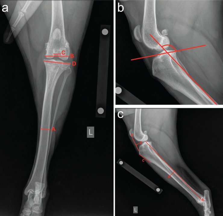
Fig 1. Illustration of radiographic measurements of dog stifles and tibiae.
Panel A. Cranial-caudal radiograph shows the mid-diaphyseal tibial width (A), the femoral condyle width (B), the femoral notch width (C), and the proximal tibial width (D). Panel B. Lateral radiograph of proximal tibia illustrating the tibial plateau angle (see Table 1 for description). Panel C. Lateral tibial radiograph illustrating the tibial length (A), the tibial diaphyseal width (B), the tibial tuberosity length (C), the infrapatellar fat pad height (D), and the infrapatellar fat pad width (E).

