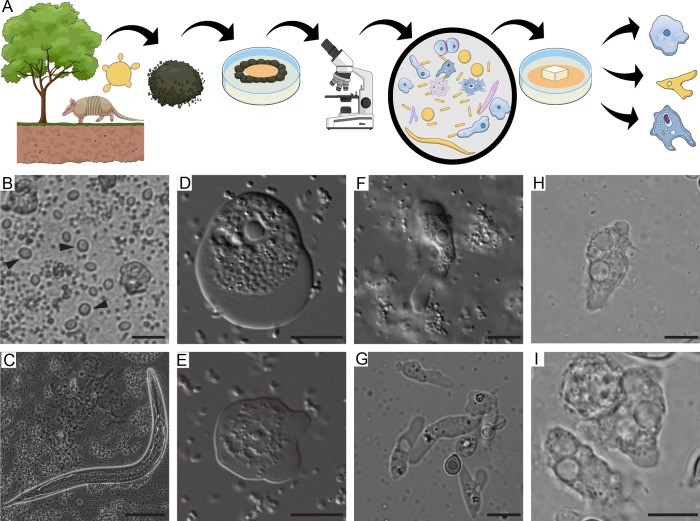
Fig 1. Soil organisms sharing the putative habitat of P. brasiliensis.
A) Schematic representation of the soil amoeba isolation methodology. Soil samples from armadillo burrows positive for P. brasiliensis DNA were collected and used for the isolation of soil amoebae. The samples were plated in non-nutrient agar plates containing a bacterial lawn as a food source and observed in an inverted microscope B) Bright field microscopy of ciliate trophozoites (black arrowheads) present in a soil sample. Scale bar = 20 μm, C) Bright field microscopy of a nematode present in the soil sample. Scale bar = 50 μm, D) and E) DIC microscopy of trophozoites of A. spelaea. Scale bar = 10 μm. F and G) DIC microscopy of trophozoites of V. vermiformis. Scale bar = 10 μm. H and I) DIC microscopy of trophozoites of Acanthamoeba spp. Scale bar = 10 μm.

