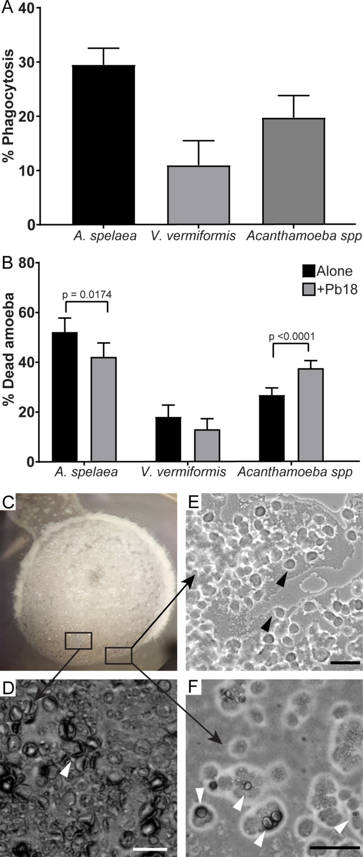
Fig 2. Interaction between P. brasiliensis Pb18 with amoebae isolated from soil of armadillo burrows positive for P. brasiliensis.

The amoeba isolates were co-incubated with Pb18 previously dyed with pHrodo or FITC at an MOI of two at 25°C for 24 hours in PYG medium. A) Percentage of amoeba cells interacting with P. brasiliensis Pb18. After the interaction, non-internalized Pb18 cells were dyed using Uvitex 2B. B) Viability of the different amoeba isolates after 24 hours of interaction with Pb18. A and B depict the results of at least three independent experiments. At least 100 cells per replicate of each sample were counted for each assay. The bars represent 95% confidence intervals. C-F) a suspension of A. spelaea cells was placed next to a colony of P. brasiliensis cells in non-nutrient agar. The cells were co-incubated at 25°C and examined daily in an inverted microscope. C) Macroscopic view of the fungal colony in a 35 mm plate. D) Microscopic view of the fungal cell lawn after seven days of interaction. E) Microscopic view of amoeba trophozoites growing in the periphery of the fungal lawn. F) Microscopic view of amoeba trophozoites interacting with a fungal cell. Scale bars = 50 μm. Black arrowheads indicate trophozoites. White arrowheads indicate fungal cells.
