Figure 3.

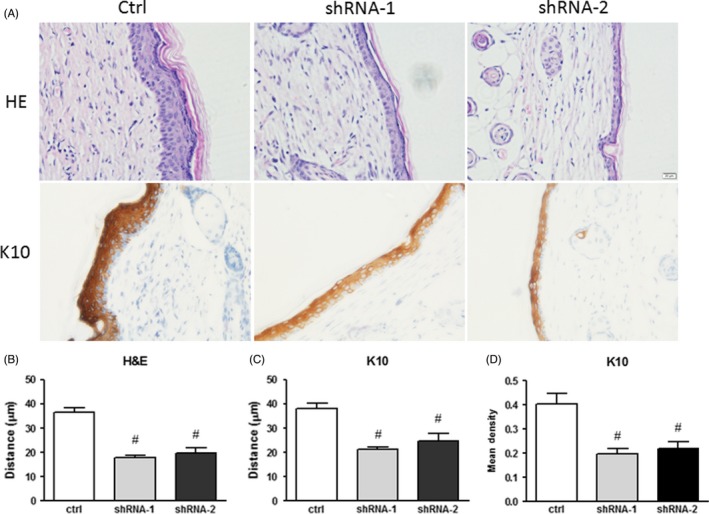
Depletion of Jarid1b impaired suprabasal layer formation. A, Representative images of H&E and K10 immunohistochemical staining in HaCaT reconstituted skin. B, Full‐thickness analysis of H&E staining in the epidermis. C, K10 staining thickness and (D) intensity analysis (#P < 0.01 shRNA group vs control at each time point, Student's t test)
