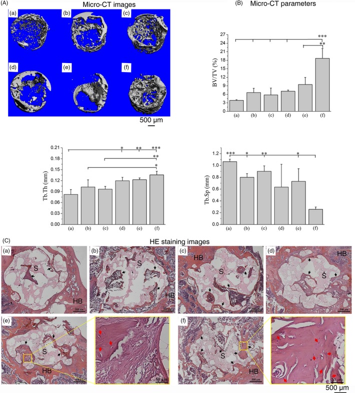
Figure 5.

A, Micro‐CT images of the regenerated bone tissue and (B) relevant bone parameters of BV/TV (bone tissue volume/total volume), Tb.Th (trabecular thickness) and Tb.Sp (trabecular separation). C, Histological evaluation (HE staining) of new bone formation within the pore at 8 wk: (a) pure scaffold, (b) MSCs scaffold, (c) OMSCs scaffold, (d) ECs scaffold, (e) MSCs/ECs (0.5/1.5) scaffold and (f) OMSCs/ECs (0.5/1.5) scaffold. HB—host bone; S—scaffold; black arrows—new bone; red arrows—capillary vessels. *P < 0.05; **P < 0.01; ***P < 0.001
