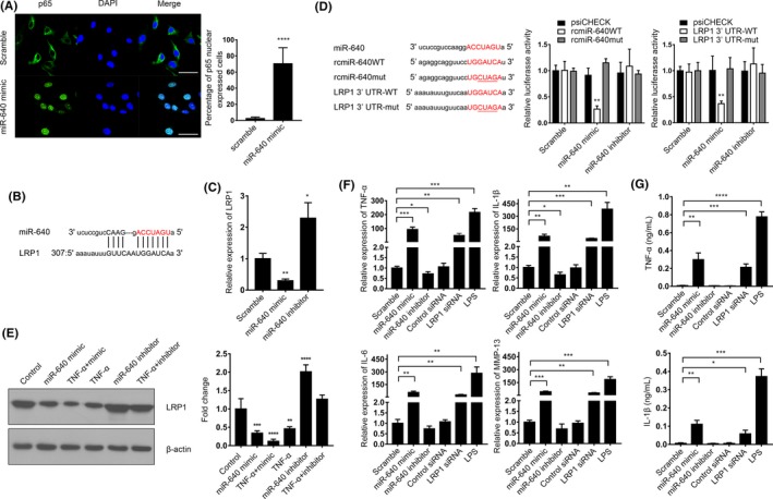
Figure 4.

miR‐640 targeted to LRP1 to enhance NF‐κB signalling pathway activity. A, The immunocytochemistry assay was performed to reveal the location of p65 in miR‐640 overexpressed NP cells. The view field was randomly selected. Scar bar, 50 µm. B, The schematic diagram of miR‐640 targeted to LRP1. C, The mRNA levels of LRP1 in NP cells with or without miR‐640 expression were measured by qPCR. D, 293T cells were transfected with the indicated plasmids and miR‐640 mimic or inhibitor. Then, relative luciferase activities were measured to verify that miR‐640 directly bound to LRP1 3’UTR and promoted its degradation. E, LRP1 expression was detected by Western blot in the miR‐640 overexpressed or inhibited NP cells combined with 24 h of TNF‐α treatment. F, The mRNA expression of the NF‐κB signalling pathway target genes was detected by qPCR in NP cells with the indicated treatments. G, The ELISA assay was performed to measure the contents of the secreted TNF‐α and IL‐1β in the medium of NP cells with the indicated treatments. One‐way ANOVA was used to evaluate the significant difference for these data. *P ˂ .05; **P ˂ .01; ***P ˂ .001; ****P ˂ .0001
