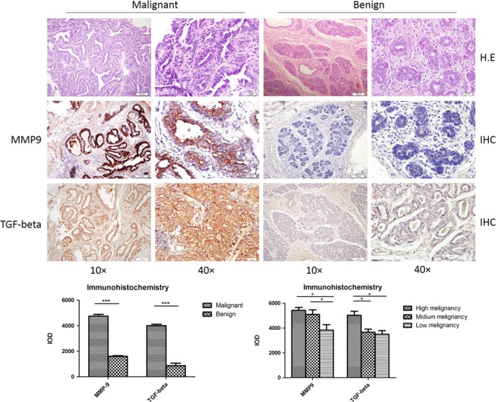
Figure 1.

Histopathological assays of canine breast malignant and benign tumours. A, Pathological assays. Upper panel: representative images of HE stain; middle panel: representative images of matrix metalloproteinase (MMP)‐9‐specific immunohistochemical (IHC). Bottom panel: representative images of transforming growth factor beta (TGF‐β)‐specific IHC. The magnifications are shown. B, Quantitative assays of the average optical densities of MMP‐9 and TGF‐β signals in the malignant and benign tumours. Statistical analysis was performed using one‐way ANOVA. ***P < 0.001 and *P < 0.05 were considered significantly different
