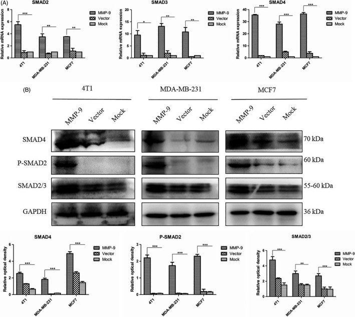
Figure 5.

Analyses of the changes in cellular SMADs in the cells transfected with pMMP‐9‐HA. Cells were harvested 48 h post‐transfection. A, qRT‐PCR assays. The total RNA was prepared, and the transcriptional levels of various SMAD genes were evaluated with the individual qRT‐PCRs. Y‐axis represents the values of 2−ΔΔCT. Each test was repeated for three times. Graphical data denote mean ± SD. B, Western blots. The cellular levels of various SMADs were determined with the individual Western blots. Quantitative assays of the relative grey value of each SMAD after normalized with the data of the individual GAPDH are shown. Each test was repeated for three times. Graphical data denote mean ± SD. Statistical analysis was performed using two‐way ANOVA. ***P < 0.001, **P < 0.01 and *P < 0.05 were considered significantly different
