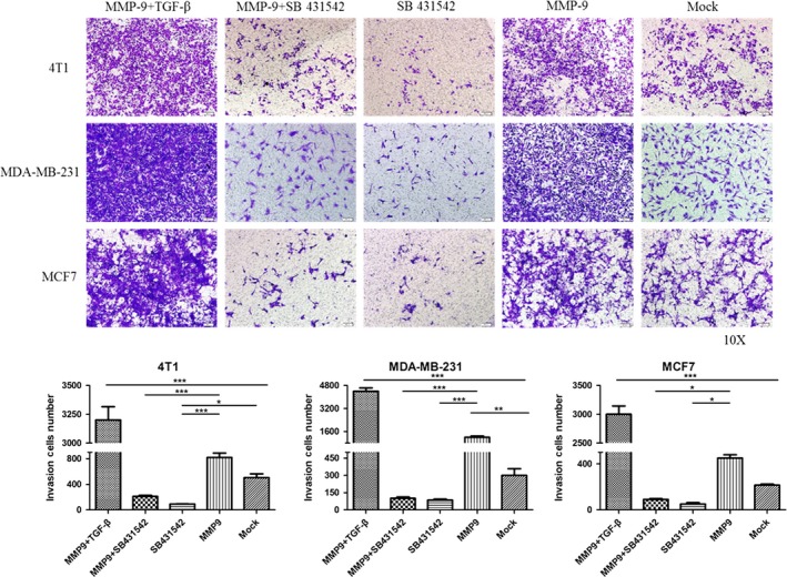
Figure 7.

Influences of the transforming growth factor beta (TGF‐β) inhibitor on the invasion abilities of the cells transfected with pMMP‐9‐HA. Representative images of cells in the lower side of the filter. Cells were exposed to recombinant TGF‐β and/or the inhibitor SB 431542 12 h post‐transfection. The migration of cells to the lower side of the filter was determined by crystal violet staining. Each testing group contained at least three independent wells. Statistical analysis was performed using one‐way ANOVA. ***P < 0.001, **P < 0.01 and *P < 0.05 were considered significantly different
