
Figure 4.
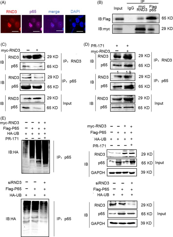
RND3 binds directly to p65 and degrades p65 protein through the ubiquitin‐mediated proteosomal pathway. A, Confocal microscopy of RND3 and p65 localizations in GBM cells (scale bar = 20 µm). B, Coimmunoprecipitation assay in U87 cells co‐transfected with a myc‐RND3 expression construct and Flag‐tagged p65 in U87 cells. C, Coimmunoprecipitation of RND3 and endogenous p65 in myc‐RND3‐transfected U87 cells and controls U87 cells transfected with vector control plasmid (myc). D, Coimmunoprecipitation of RND3 and endogenous p65 in myc‐RND3‐ and control‐transfected U87 cells treated with PR‐171 (20 nm). E, Representative immunoblots of p65 and ubiquitinated p65 in U87 cells when RND3 was up‐ or downregulated and PR‐171 was used to treat U87 cells. IP: specific antibody used to pulled down proteins; IB: specific antibody used to detected protein expression level by Western blot