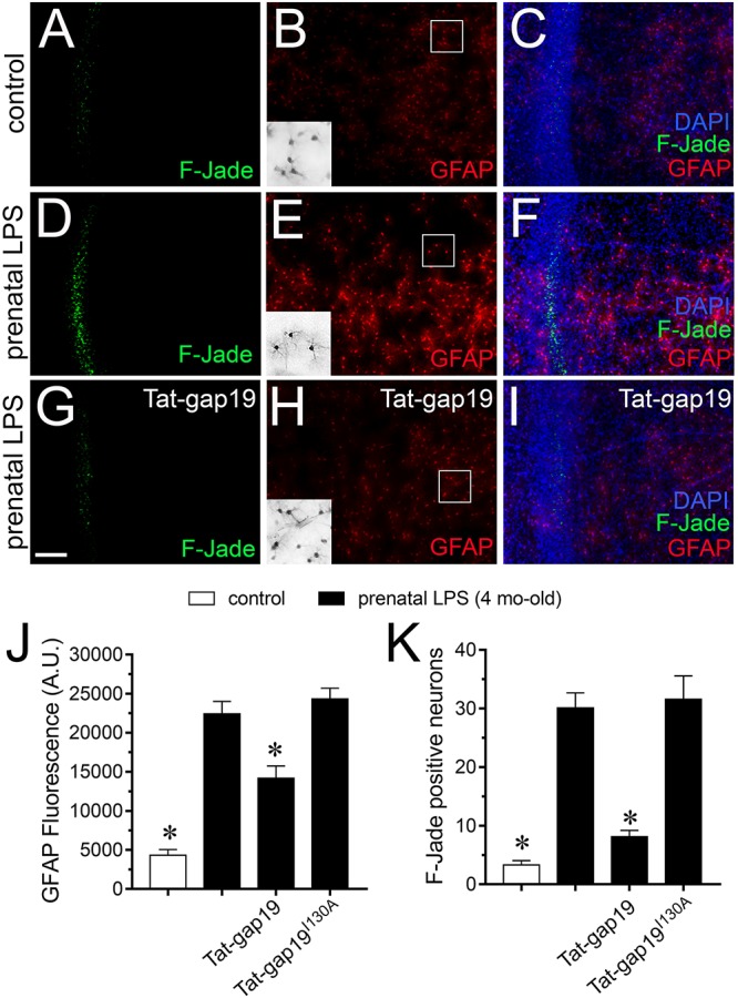
FIGURE 8.

Cx43 hemichannel contributes to neuronal death evoked by prenatal LPS exposure on offspring hippocampus. (A–I) Representative images depicting Fluoro-Jade (F-Jade, green), GFAP (red) and DAPI (blue) staining in acute slices from control offspring (A–C) or prenatally LPS-exposed offspring of 4 months old alone (D–F) or in combination with the in vivo administration of 23 mg/kg Tat-gap19 (G–I). Insets of gray scale GFAP staining for astrocytes are shown from the area depicted within the white squares in (B,E,H). (J) Averaged data of GFAP fluorescence per field in acute slices from control offspring (white bars) or prenatally LPS-exposed offspring of 4 months old alone (black bars) or in combination with the in vivo administration of 23 mg/kg Tat-gap19 (gray bars). ∗p < 0.01 versus LPS, one-way ANOVA Tukey’s post hoc test, mean ± S.E.M., n = 3. (K) Averaged number of F-Jade-positive CA1 pyramidal neurons per field in acute slices from control offspring (white bars) or prenatally LPS-exposed offspring of 4 months old alone (black bars) or in combination with the in vivo administration of 23 mg/kg Tat-gap19 (gray bars). ∗p < 0.001 versus LPS, one-way ANOVA Tukey’s post hoc test, mean ± S.E.M., n = 3. Calibration bar = 100 μm.
