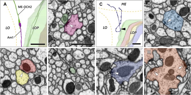
FIGURE 5.

Axon and synaptic ultrastructure in the chiasm. (A) An Am1 branch and a ME-OCH2 cell terminal, both terminating in OCH2. (B) The two cells shown in (A) form a synaptic contact within OCH2. The two sites of synaptic contact are revealed where the Am1 terminal (red), forms a T-bar ribbon (arrowhead), which is presynaptic, opposite the ME-OCH2 terminal (green) where it is postsynaptic. (C) Part of a DCV cell making a reverse turn in OCH2. Its ME part is incompletely reconstructed. (D) A synaptic site of the DCV cell shown in (C). The synaptic site corresponds to the point indicated by the arrowhead in (C). The DCV cell has a presynaptic T-bar (arrowhead) and provides input to a dyad of postsynaptic Tm cell axons. (E,F) Putative septate or adherens junctions in OCH2. (E) is a case between Tm cell axons, and (F) is a case between a Tm cell axon and glia. (G,H) Typical presynaptic T-bars found outside OCH2. (G) is an example of a Tm1 cell, while (H) is a Tm9 cell terminal, both in the first stratum of the lobula. Scale bar: (A,C): 10 μm; (B), (D–H): 1 μm.
