Fig. 4.
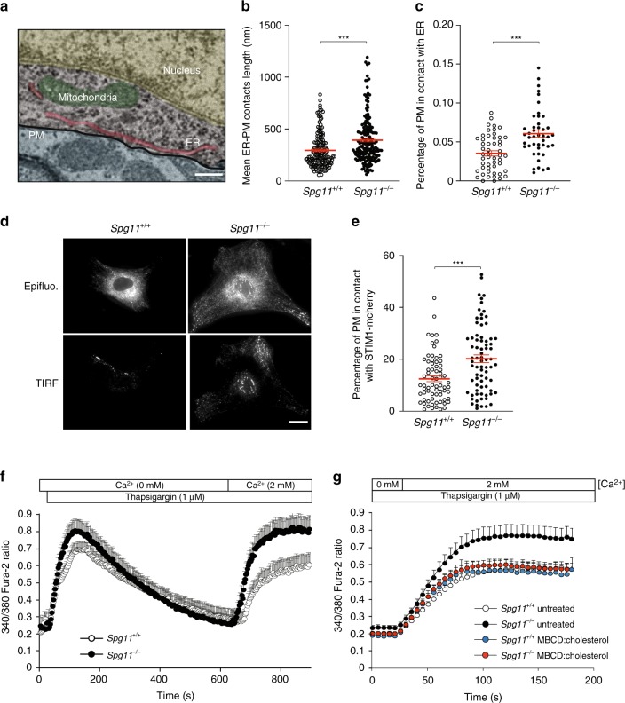
The depletion of plasma membrane cholesterol promotes higher store-operated calcium entry. a Electron micrograph of neurons in the cortex of a 2-month-old Spg11−/− mouse, showing close contact between the endoplasmic reticulum (ER) and plasma membrane (PM). False colors highlight the various cellular compartments. Scale bar: 250 nm. b, c Quantification of contacts between the ER and plasma membrane, defined as the zone where the distance between the two membranes is lower than 30 nm. b Quantification of the mean length of individual contacts between the ER and plasma membrane in the cortex of 2-month-old Spg11−/− or Spg11+/+ mice. c Quantification of the percentage of the plasma membrane in close contact with the ER in the cortex of 2-month-old Spg11−/− or Spg11+/+ mice. The graphs represent the mean ± SEM. N > 23 cells analyzed in two independent mice for each group. T-test: ***p < 0.0001. d Spg11−/− or Spg11+/+ mouse embryonic fibroblasts transfected with vectors expressing STIM1-mCherry imaged by epifluorescence or total internal reflection microscopy (TIRF). Scale bar: 10 µm. e Quantification of the percentage of the cellular area with STIM1-mCherry staining detected by TIRF microscopy, indicating close contact between STIM1-mCherry and the plasma membrane. The graph shows the mean ± SEM. N > 60 cells from three independent experiments. T-test: ***p < 0.0001. f Evaluation of extracellular calcium import by SOCE. Cytosolic calcium was measured with Fura-2 in the absence of extracellular calcium. The ER calcium store was depleted with thapsigargin, 2 mM CaCl2 added to the extracellular medium, and the increase in cytosolic calcium measured with Fura-2, allowing the quantification of SOCE. The graph shows the mean ± SEM. N > 35 cells from three independent experiments. g Increasing cholesterol levels in the plasma membrane with methyl-β-cyclodextrin (MBCD) loaded with cholesterol decreases store-operated calcium entry in Spg11−/− fibroblasts, measured by the addition of 2 mM extracellular calcium after a 10-min treatment with thapsigargin. The graph shows the mean ± SEM. N > 60 cells from three independent experiments

