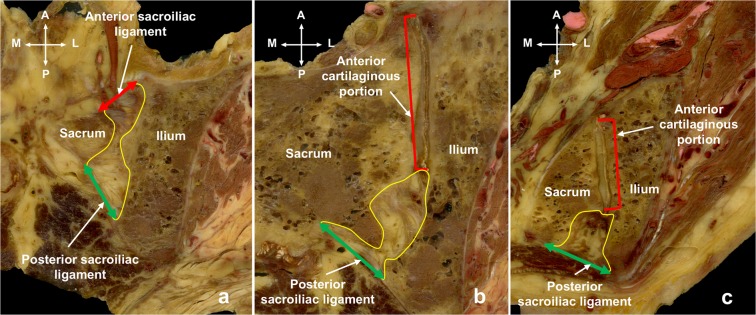
Figure 2.
(a) A slice from the superior sub-region of the posterior sacroiliac joint. Boundaries of the posterior sacroiliac joint (in yellow) at the level of the first sacral body in horizontal images. Laterally and medially the region is delimitated by the iliac and sacral bones. It is deep to the posterior sacroiliac ligament (green) posteriorly and deep to anterior cartilaginous portion of the sacroiliac joint (red) anteriorly. (b) A slice from the middle sub-region of the posterior sacroiliac joint. Boundaries of the posterior sacroiliac joint (in yellow) at the level of the second and third sacral bodies. Laterally and medially the region is delimitated by the iliac and sacral bones. It is deep to the posterior sacroiliac ligament (green) posteriorly and deep to the anterior articular cartilaginous portion (red) anteriorly. (c) A slice from the inferior sub-region of the posterior sacroiliac joint. Boundaries of the posterior sacroiliac joint (in yellow) at the level of the third and fourth sacral bodies. Laterally and medially the region is delimitated by the iliac and sacral bones. It is deep to the posterior sacroiliac ligament (green) posteriorly and deep to the anterior articular cartilaginous portion (red) anteriorly. A: anterior, L: lateral, M: medial, P: posterior.

