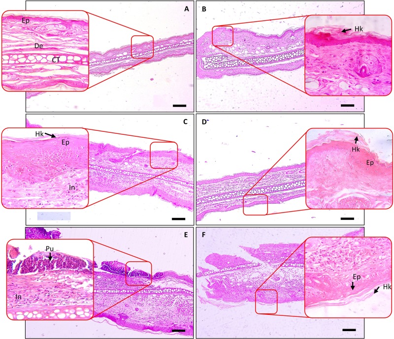
Figure 6.
Histopathological analysis of SBKT treatment on TPA-induced ear psoriasis in mice. Histopathological analysis of mice ear tissue was performed following fixation and hematoxylin and eosin staining. Low-magnification images were obtained at 100×, and the higher-magnification image was obtained at 400×. (A) Normal control: represents normal epidermis (Ep), dermis (De), sebaceous gland (Sg), cartilage (CT). (B) Vehicle control (acetone) treated ear: represents normal epidermis (Ep), dermis (De), sebaceous gland (Sg), cartilage (CT). (C) TPA-CON: represents hyperkeratosis (Hk) and hyperplastic epidermis (Ep), presence of inflammatory cells (In) in the dermis region. (D) TPA and DEXA (0.2 mg/ear) treated ear: reduced hyperplastic epidermis (Ep), absence of inflammatory cells in the dermis region. (E) TPA and SBKT (100 mg/kg; p.o. and 20 μl; T.A.) treated ear: reduced hyperkeratosis (Hk) and hyperplastic epidermis (Ep), reduced presence of inflammatory cells (In) in the dermis region. (F) TPA and SBKT (200 mg/kg; p.o. and 20 μl; T.A.) treated ear: reduced hyperkeratosis (Hk) and hyperplastic epidermis (Ep). The scale represents 100 µm (n = 8 animals).

