
Figure 5.
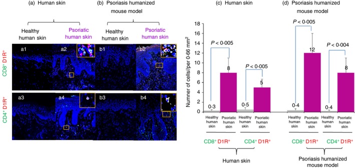
Immunofluorescence analysis of T cells expressing D1R in human psoriatic skin, and in human psoriasiform skin grafts of Psoriasis humanized mouse model in SCID/Beige mice. (a) CD8+/D1R+ and CD4+/D1R+ T cells are more numerous in involved skin from patients with Psoriasis than in healthy skin. (b) These CD8+/D1R+ and CD4+/D1R+ double‐positive T cells are also elevated in psoriatic human skin of Psoriasis humanized mouse model, compared with healthy human skin. (c) Quantitative analysis of double‐positive CD8+/D1R+ and CD4+/D1R+ T cells in human Psoriasis and in (d) psoriatic and healthy skin of Psoriasis humanized mouse model. The data represent mean + SD of six Psoriasis patients. Statistical analysis used the Kruskal–Wallis test. Statistical significance was set at P < 0·05.