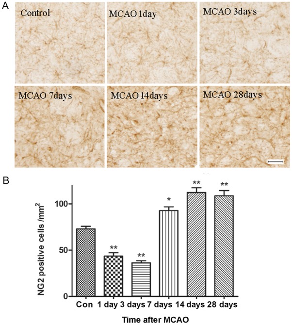
Figure 4.
Immunostaining of OPCs in the ischemic area. (A) Representative images of sections stained with anti-NG2, a marker of OPCs, at day 1, 3, 7, 14 and 28 after reperfusion. Scale bar, 50 µm. There was a significant increase in the number of OPCs and this was accompanied by morphological changes in these cells from day 7 to 28 after reperfusion. (B) Statistical analysis of the number of NG2 positive cells. n=6. *P<0.05, **P<0.01 vs. Con. MCAO, middle cerebral artery occlusion; Con, control; OPCs, oligodendrocyte precursor cells; NG2, chondroitin sulfate proteoglycan 4.

