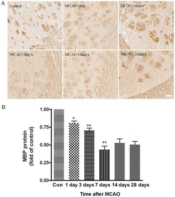
Figure 5.
Measurement of MBP expression in the ischemic brain tissues using immunohistochemistry. (A) Representative images of sections stained with anti-MBP, a marker for myelin, after MCAO. Scale bar, 50 µm. (B) Quantification of the average intensity as shown in (A). The intensity of MBP decreased from day 3 to 28 after MCAO. n=6. *P<0.05, **P<0.01 vs. Con. MBP, myelin basic protein; MCAO, middle cerebral artery occlusion; Con, control.

