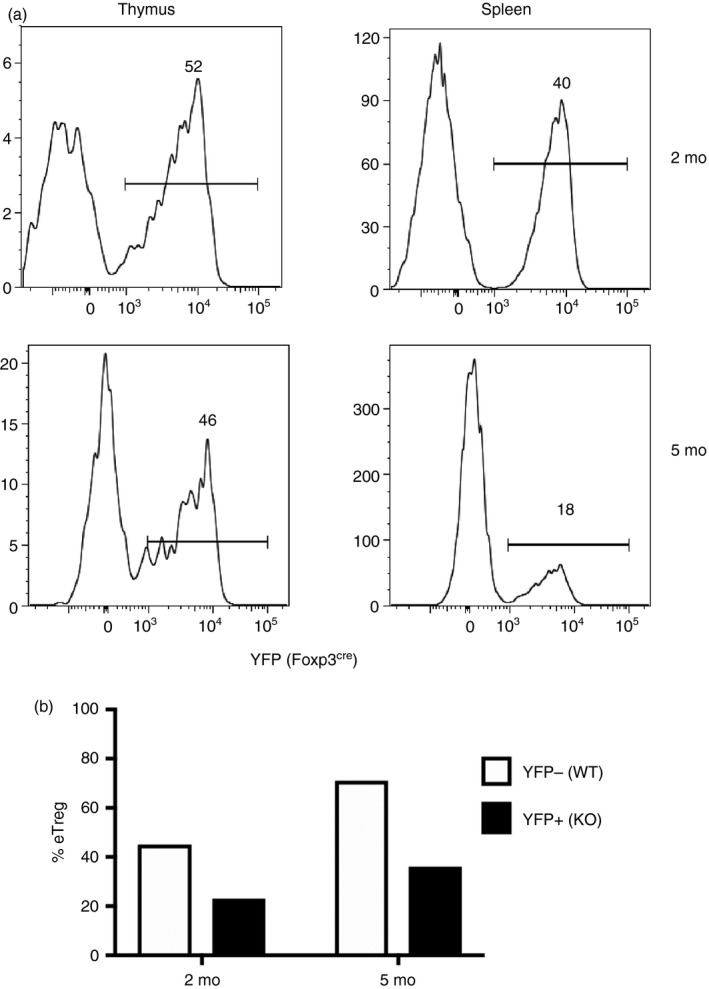
Figure 3.

Analysis of heterozygous Foxp3 females. (a) CD4+ Foxp3+ thymocytes or splenocytes from 2‐ or 5‐month‐old Ikzf2fl/fl × Foxp3wt/ YFP ‐Cre mice were analyzed by flow cytometry for YFP expression, a reporter for the Helios‐deficient cells. (b) YFP − (wild‐type; WT) or YFP + (knockout; KO) CD4+ Foxp3+ Treg cells were analyzed for CD44 and CD62L to calculate the percent of eTreg cells defined as CD44+ CD62L−.
