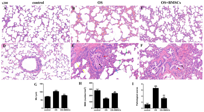
Figure 3.
The therapeutic effects of BMSCs on OS model rats. Hematoxylin and eosin (H&E) staining of lung tissue sections in the control group (A and D), OS group (B and E) and OS+BMSC group (C and F). Scale bar, 50 µm. (A) Alveolar structure was intact. (B) Alveolar size was different, the alveolar number was decreased, the alveolar space was enlarged and the alveolar septum was fractured. (C) Less enlarged alveolar space and less alveolar walls were thickened. (D) Alveolar structure was intact, and no inflammatory cell infiltration was observed. (E) A small number of bronchial epithelial cells were shed, and the bronchial epithelial cells exhibited edema and degeneration; the bronchial tubes were irregular in shape and narrow in the lumen; alveolar walls were thickened; the alveoli fused into a large cystic cavity; and much inflammatory cell infiltration. (F) Decreased inflammatory cell infiltration. Histological assessment of the sections was determined using the MLI (G), MAN (H) and pathological scores (I). A small number of bronchial epithelial cells are shed in the field, as shown by the black arrow. More bronchial epithelial cells showed edema and degeneration, cell swelling, loose cytoplasm and light staining, as shown by red arrows. Thickening of the alveolar walls is noted in the field, as shown by yellow arrows. The alveoli fused into a large cystic cavity as shown by green arrows. Inflammatory cells infiltrate the alveolar walls as shown by a blue arrow. ***P<0.001 vs. control group; ##P<0.01 and ###P<0.001 vs. OS group. BMSCs, bone marrow-derived mesenchymal stem cells; OS, overlap syndrome; MLI, mean linear intercept; MAN, mean alveolar number.

