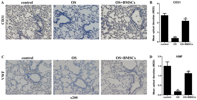
Figure 5.
The effects of BMSCs on CD31 and VWF levels in alveolar walls in the OS+BMSC group rats. (A and C) The expression levels of CD31 and VWF were detected by an immunohistochemical assay (magnification, ×400). Scale bar, 50 µm. (A) Brown staining of cells in the control group. A small amount of brown was observed in the alveolar wall of the OS group, and increasing brown staining of cells in the OS+BMSCs group. (B) Quantitative results of the CD31 protein immunohistochemical assay. (C) A large number of stained cells was observed in the control group, less brown was observed in the alveolar wall of the OS group, and increased brown staining of cells was observed in the OS+BMSCs group. (D) Quantitative results of the VWF protein immunohistochemical assay. The brown color shows the positive cells for CD31 and VWF. The blue color shows the nuclei. **P<0.01 and ***P<0.001 vs. control group; ##P<0.001, vs. OS group.

