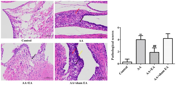
Figure 3.
Effects of EA on ankle joint damage in AA rats. Representative microphotographs of ankle joint sections from various groups (H&E stain, magnification, ×400). Data are expressed as the mean ± standard deviation (n=10). **P<0.01 vs. the control group; ##P<0.01 vs. the AA group. The red arrows indicate hyperplastic synoviocytes, the black arrows indicate infiltrating inflammatory cells, and the yellow arrows indicate hyperplastic synovial lining cells. EA, electroacupuncture; AA, adjuvant arthritis; H&E, hematoxylin and eosin.

