Figure 1.
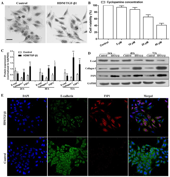
HDM combined with TGF-β1 induces EMT in HBECs. (A) HDM combined with TGF-β1 induced a morphological phenotype that was characteristic of EMT. Scale bar, 20 µm. (B) Cyclopamine treatment was shown to lead to the inhibition of 16HBEC survival in a dose-dependent manner using a CCK-8 assay. (C and D) Cells (16HBECs) were stimulated with HDM (30 µg/ml) and TGF-β1 (5 ng/ml) or in complete medium for 24–72 h. Western blotting demonstrated that HDM/TGF-β1 treatment increased the expression of mesenchymal markers (collagen I and FSP1) and reduced the expression of an epithelial marker (E-cadherin). An antibody against GAPDH was used as an internal control. *P<0.05 and **P<0.01 vs. the control. (E) As shown by immunofluorescence staining, the expression of E-cadherin was decreased in the 16HBECs after 72 h of treatment with HDM/TGF-β1, and the expression of FSP1 increased. Magnification, ×400. HDM, house dust mite; TGF-β1, transforming growth factor β1; EMT, epithelial-mesenchymal transition; HBECs, human bronchial epithelial cells.

