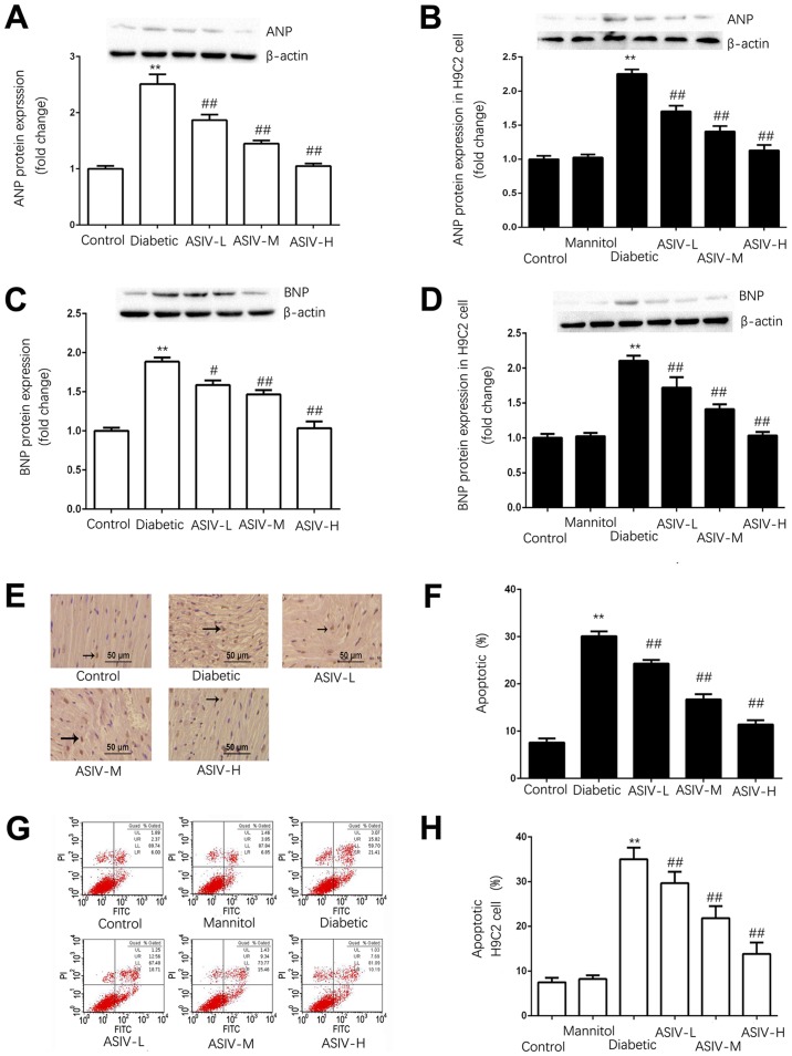
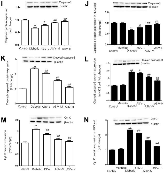
Figure 3.
ASIV decreases the damage of myocardial tissue of diabetic rats and H9C2 cells in high-glucose environment. Protein expression of ANP in (A) the myocardium and (B) H9C2 cells. The protein expression of BNP in (C) the myocardium and (D) H9C2 cells. (E) Myocardial apoptosis assayed by TUNEL staining. TUNEL-positive cells were manifested as a marked appearance of dark brown apoptotic cell nuclei, and (F) the percentage of apoptotic cells in myocardial tissue. (G) The detection of apoptosis in H9C2 cells by AnnexinV-FITC and PI double staining, and (H) the percentage of apoptotic H9C2 cells. The protein expression of (I) caspase-3, (J) cleaved caspase-3 and (K) cytoplasmic Cyt C in the myocardium, and (L) caspase-3, (M) cleaved caspase-3 and (N) cytoplasmic Cyt C in H9C2 cells. The data are expressed as the means ± SD. n=4. **P<0.01 vs. control group. #P<0.05 and ##P<0.01 vs. diabetic group. The black arrows indicate the nucleus of apoptotic cardiomyocytes. ANP, atrial natriuretic peptide; ASIV, astragaloside IV; L, low; M, mid; H, high; BNP, brain natriuretic peptide; PI, propidium iodide; Cyt C, cytochrome c.


