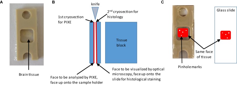
FIGURE 2.
Procedure for the preparation of adjacent brain cryo-sections. Needle holes orientation marks are made around the region of interest on the tissue block before sectioning. (A) A first brain cryo-section is deposited and maintained between two polycarbonate films stretched over a PEEK sample holder. This first cryo-section will be used for metal analysis. (B) Explanation of how to keep the tissue orientation for correlative imaging. The second cryo-section is used for immunohistochemistry. This second cryo-section is deposited on a glass microscopy slide with the face adjacent to the first one on the top. (C) Illustration of how to identify regions of interest on both sections thanks to the needle holes orientation marks.

