FIGURE 3.

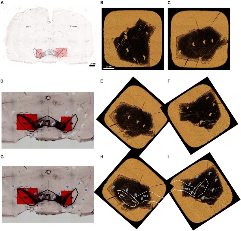
Overall procedure for superposition of TH immuno-histochemical sections and the adjacent freeze dried unstained section for PIXE analysis. (A) Entire brain section stained with TH to localize the dopaminergic pathway, SNpc, SNpr, and VTA. (B,C) Optical transmission microscopy images of freeze dried sections for PIXE analyses (B: IpsiL brain side and C: ContraL brain side). (D,G) Zoom of the stained region, to distinguish the pinholes done around the SN (indicated with blue stars). These images have a horizontal flip with respect to the initial image in order to obtain the same orientation for the pinholes than on freeze dried samples. (E,F) PIXE section images, applying a rotation, to display the same orientation of pinholes than on the immuno-histochemical sections. (H,I) Correctly oriented images using the pinholes (marked with a blue star) to locate precisely the SNpc, SNpr, and VTA regions on the sections for PIXE analysis. Scale bars = 1 mm.
