FIG 1.
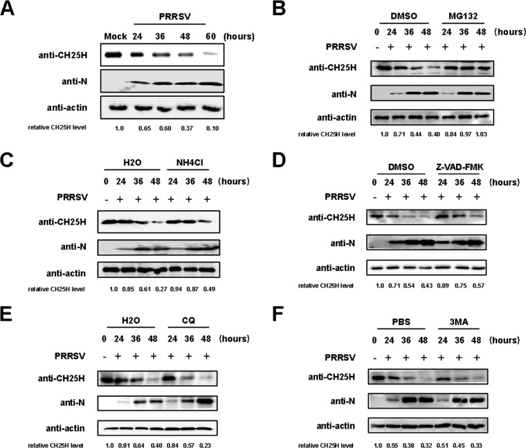
The ubiquitin-proteasome pathway plays a major role in the degradation of pCH25H during PRRSV infection. (A) PAMs were infected with PRRSV (MOI of 1.0). At various time points (24, 36, 48, and 60 h) postinfection, cells were harvested and pCH25H expression was analyzed by Western blotting. (B to F) PAMs were infected with PRRSV (MOI of 1.0) and treated with various inhibitors, including MG132 (10 μM) (B), NH4Cl (10 mM) (C), Z-VAD-FMK (10 μM) (D), CQ (20 μM) (E), or 3-MA (5 mM) (F). At 24, 36, and 48 h postinfection, cells were harvested and pCH25H expression was analyzed by Western blotting. ImageJ software was used to analyze the relative levels of pCH25H in comparison with mock-infected cells, and the ratios are displayed as fold changes below the images. DMSO, dimethyl sulfoxide.

