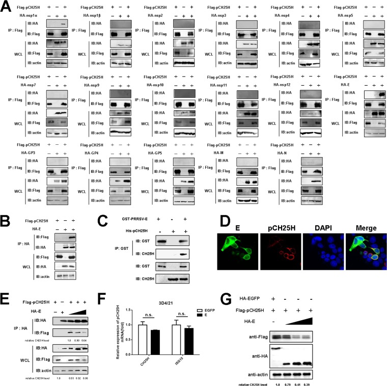
FIG 2.
PRRSV E protein interacts with pCH25H and downregulates its expression. (A) HEK-293T cells were cotransfected with expression vectors encoding FLAG-tagged pCH25H and HA-tagged PRRSV proteins. The cells were lysed at 48 h after transfection and immunoprecipitated with anti-FLAG antibody. Whole-cell lysate (WCL) and immunoprecipitation (IP) complexes were analyzed by immunoblotting (IB) with anti-FLAG, anti-HA, or anti-β-actin antibodies. (B) HEK-293T cells were cotransfected with expression vectors encoding HA-tagged E protein and FLAG-tagged pCH25H. The cells were lysed at 48 h after transfection and immunoprecipitated with an anti-HA antibody. Whole-cell lysate and immunoprecipitation complexes were analyzed by immunoblotting with anti-FLAG, anti-HA, or anti-β-actin antibodies. (C) The purified GST-E protein was incubated with glutathione-Sepharose 4B beads, and then the recombinant protein His-pCH25H was added. After being washed three times, the beads were collected for Western blotting with antibodies against GST or CH25H. (D) HEK-293T cells were cotransfected with FLAG-tagged pCH25H and HA-tagged E protein expression vectors. At 48 h posttransfection, cells were fixed for immunofluorescence assays for pCH25H and E proteins using primary antibodies (rabbit anti-HA and mouse anti-FLAG) followed by secondary antibodies (AF594-conjugated donkey anti-mouse IgG and AF488-conjugated donkey anti-rabbit IgG). Nuclei were counterstained with DAPI. Fluorescence images were acquired with a confocal laser scanning microscope (Olympus FluoView v3.1). (E) HEK-293T cells were cotransfected with expression vectors encoding FLAG-tagged pCH25H and different doses of HA-tagged PRRSV E protein. The cells were lysed at 48 h after transfection and immunoprecipitated with an anti-HA antibody. (F) 3D4/21 cells were cotransfected with expression vectors encoding FLAG-tagged pCH25H and HA-tagged PRRSV E protein. At 48 h posttransfection, cells were harvested for quantitation of pCH25H mRNA by qRT-PCR. Experiments were performed in triplicate, and data are representative of three independent experiments. The standard deviations are indicated by error bars. (G) 3D4/21 cells were cotransfected with an expression vector encoding FLAG-tagged pCH25H and increasing amounts of an expression vector encoding HA-tagged E protein. At 48 h posttransfection, expression of FLAG-pCH25H and HA-E protein was analyzed by Western blotting. The relative levels of pCH25H in comparison with HA-EGFP-transfected cells were analyzed by ImageJ software, and the ratios are displayed as fold changes below the images. n.s., not significant.

