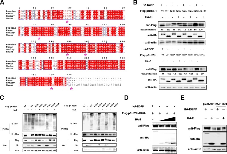
FIG 4.
The Lys28 site of pCH25H is ubiquitinated by PRRSV E protein. (A) CH25H sequences from different species were aligned (*, lysine). (B) 3D4/21 cells were cotransfected with expression vectors encoding FLAG-tagged wild-type (WT) pCH25H or pCH25H mutants (K28A, K72A, K148A, K158A, K259A, or K270A) and HA-tagged PRRSV E protein. At 48 h posttransfection, cells were harvested for analysis of pCH25H expression by Western blotting. The numbers below the images represent the relative levels of pCH25H, compared to that of the corresponding control group, as determined via ImageJ analysis. (C) HEK-293T cells were cotransfected with expression vectors encoding HA-tagged PRRSV E protein and FLAG-tagged wild-type pCH25H or pCH25H mutants (K28A, K72A, K148A, K158A, K259A, or K270A). The cells were lysed at 48 h after transfection and immunoprecipitated with an anti-FLAG antibody. Whole-cell lysate (WCL) and immunoprecipitation (IP) complexes were analyzed by immunoblotting (IB) with anti-FLAG, anti-HA, anti-ubiquitin (Ub), or anti-β-actin antibodies. (D) 3D4/21 cells were cotransfected with expression vectors encoding FLAG-tagged K28A and different amounts of expression vectors encoding HA-tagged E protein. At 48 h posttransfection, cells were harvested for analysis of pCH25H expression by Western blotting. (E) 3D4/21 cells were cotransfected with expression vectors encoding FLAG-tagged pCH25H or hCH25H and HA-tagged E protein. At 48 h posttransfection, cells were harvested for analysis of pCH25H expression by Western blotting.

