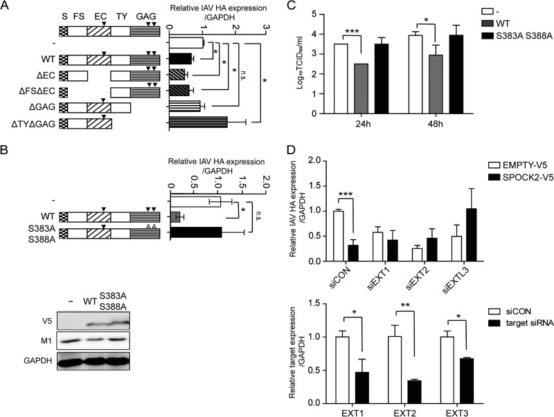
FIG 4.
The heparan sulfate moiety is responsible for the antiviral effect of SPOCK2. (A) Domain deletion mutants of SPOCK2 were overexpressed in A549 cells and infected with PR8 IAV for 24 h. HA vRNA levels were measured by qRT-PCR. (B) Site-directed mutants of SPOCK2 were overexpressed in A549 cells and infected with PR8 IAV for 24 h. HA vRNA levels were measured by qRT-PCR. For the bottom portion, cell lysates from the top portion were analyzed with anti-M1 and anti-V5, and anti-GAPDH was detected as a loading control. (C) At 36 h posttransfection, A549 cells were infected with IAV at an MOI of 1 per well for 24 h or 48 h. The supernatants from cells infected with A549 cells were collected, and the viral titers were measured by the TCID50 method. The bars indicate the mean values ± SDs obtained from three experiments. (D) A549 cells were transfected with EXT1, -2, and -3 siRNAs and a V5-tagged SPOCK2-expressing construct or empty vector. HA vRNA levels were measured by qRT-PCR. The expression was normalized by GAPDH (upper graph). The silencing efficiencies of EXT1, EXT2, and EXTL3 were measured by qRT-PCR (lower graph). Expression was normalized by GAPDH. All graphs indicate the average values ± SDs obtained from triplicate experiments. ***, P < 0.001 (t test).

