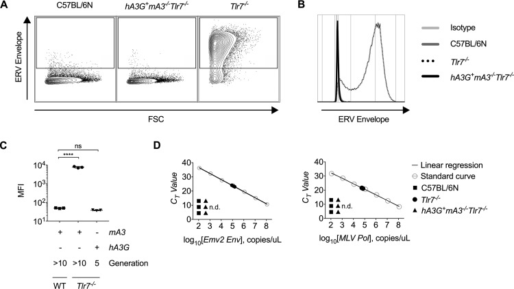
FIG 3.
Infectious ERV cannot be detected in the plasma or isolated through splenocyte coculture from hA3G+ mA3−/− Tlr7−/− mice. (A to C) Representative flow cytometry plots (A), histograms (B), and calculated mean fluorescence intensities (C) of ERV envelope expression on live, CD45.2-negative DFJ8 cells after 7 days of coculture with C57BL/6N, Tlr7−/−, or F5 hA3G+ mA3−/− Tlr7−/− splenocytes (n = 3 mice per group). (D) Absolute quantification of the number of polymerase or unspliced ERV envelope RNA copies per microliter of cDNA generated from plasma of 16-week-old C57BL/6N, Tlr7−/−, and hA3G+ mA3−/− Tlr7−/− mice. Plots are representative of three independent experiments. P values in Fig. 1C were calculated using one-way ANOVA with Dunnett’s multiple-comparison test comparing values to those of the WT control. ****, P < 0.0001; ns, not significant.

