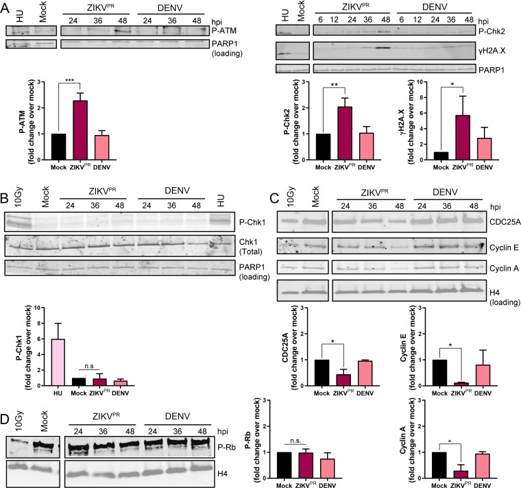
FIG 2.
ZIKV infection activates the ATM/Chk2 checkpoint in hNPCs. (A to C) Representative Western blot images and quantifications of DNA damage response signaling pathway protein expression in hNPCs infected with ZIKVPR (MOI of 0.4) or DENV (MOI of 0.4) analyzed over the time course shown (hours postinfection [hpi]). Positive controls include cells treated with 1 mM hydroxyurea (HU) for 22 h and 10-Gy-irradiated cells. Quantifications are of 48-h time points. Error bars are mean ± SD, representing the average from (A and B) three or (C) two biological replicates. (D) Representative Western blot image and quantification of phosphorylated Rb in hNPCs infected with ZIKVPR (MOI of 0.4) or DENV (MOI of 0.4) analyzed over the time course shown (hpi). The positive control is 10-Gy-irradiated cells. Quantifications are of 48-h time points. Error bars are mean ± SD, representing the average from three biological replicates. In panels A to D, * indicates P ≤ 0.05, ** indicates P ≤ 0.01, and *** indicates P ≤ 0.001 (one-way ANOVA).

