Fig. 4.
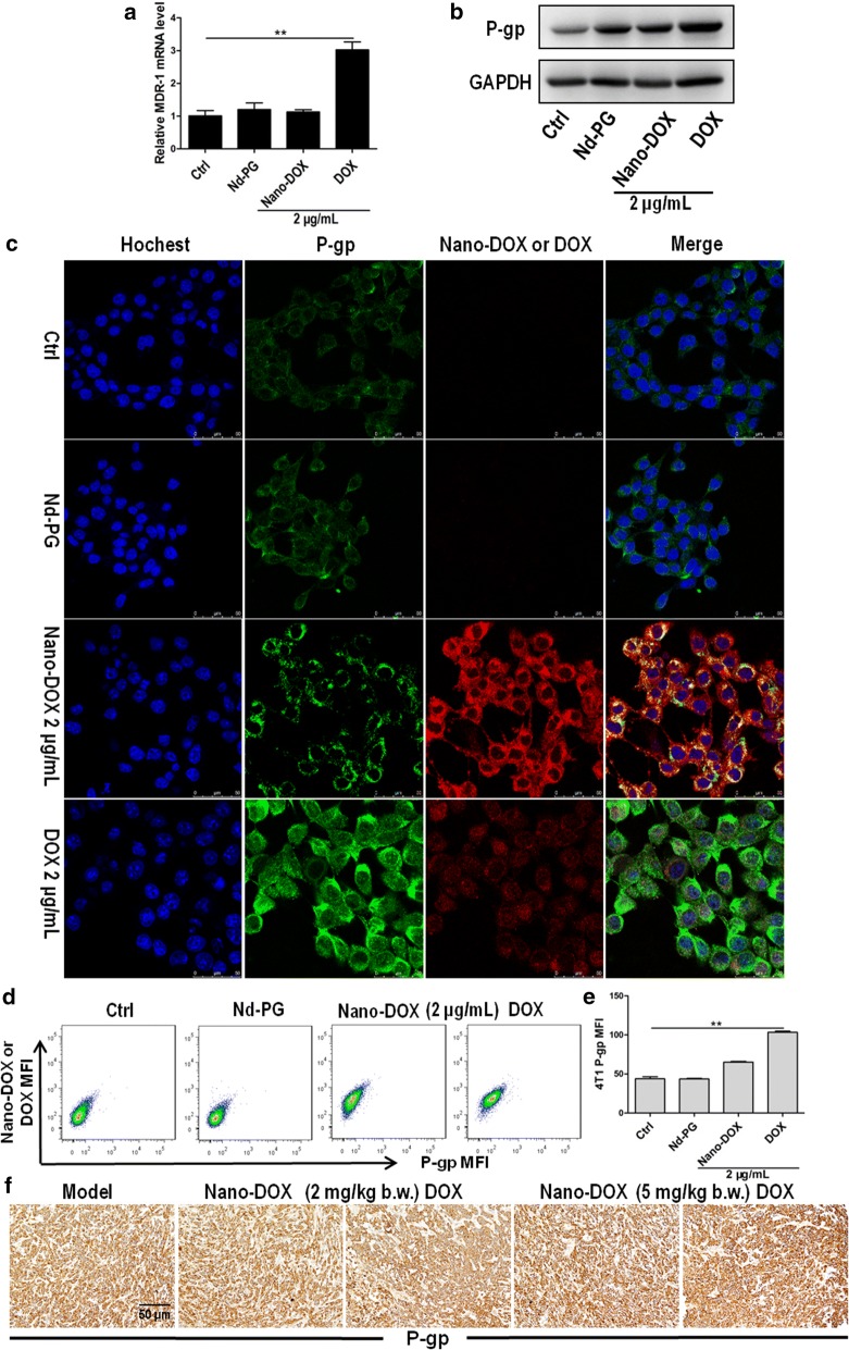
Effects of Nano-DOX and DOX on the expression of P-gp (MDR-1 protein) in 4T1 cells. a RT-PCR analysis of MDR-1 mRNA levels in in vitro 4T1 cells treated with polyglycerol-functionalized nanodiaminds (Nd-PG), Nano-DOX and DOX. b Western blotting analysis of P-gp expression in in vitro 4T1 cells treated with polyglycerol-functionalized nanodiaminds (Nd-PG), Nano-DOX and DOX. c Confocal microscopy of P-gp immunofluorescent staining in in vitro 4T1 cells treated with polyglycerol-functionalized nanodiaminds (Nd-PG), Nano-DOX and DOX. Blue fluorescence was nuclear staining by Hoechst 33342; green fluorescence was P-gp staining and red fluorescence came from Nano-DOX or DOX. d, e FACS analysis of P-gp immunofluorescent staining in in vitro 4T1 cells treated with polyglycerol-functionalized nanodiaminds (Nd-PG), Nano-DOX and DOX. f Immunohistochemical staining of P-gp in mouse orthotopic 4T1 tumor xenografts at the end of 3-week treatment of Nano-DOX or DOX. Duration of treatment was 24 h for the in vitro cell experiments. In FACS analysis, geometric means were used to quantify fluorescence intensity. Values were mean ± SD (n = 3, *p < 0.05, **p < 0.01)

