Fig. 3.
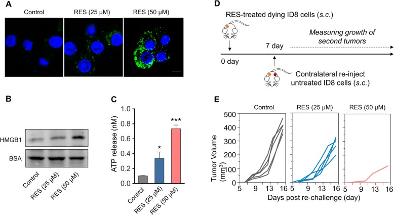
RES induces ICD in murine ovarian carcinoma cells ID8. a Fluorescent imaging of CRT exposed on surface of ID8 tumor cells after treated with RES (25 μM or 50 μM) for 24 h, and then cells were incubated with FITC-conjugated anti-calreticulin (FITC-anti-CRT) antibody for 2 h at 4 °C. After staining with DAPI, the cells were observed under confocal microscopy. b Released HMGB1 in the medium supernatant of ID8 cells treated with RES (25 μM or 50 μM) was measured by western blot, and BSA was used as the loading control. c Amount of released ATP in the medium supernatant of ID8 cells after RES treatment (25 μM or 50 μM) was determined by a chemiluminescent ATP Determination Kit. Data represent means ± SD. *p < 0.05, ***p < 0.001 (versus Control group). d Animal vaccination, using 2 rounds of subcutaneous (s.c.) injection of PBS or RES-treated 5*106 dying ID cells 7 days apart, followed by s.c. injection of 5*106 live cells on the contralateral side. Tumors growth were measured. e Growth of second tumors in animals vaccinated by dying tumor cells treated with RES or PBS

