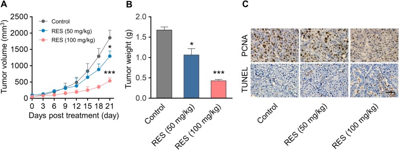
Fig. 4.
In vivo anti-tumor effect of RES in ID8 tumor model. a Growth curve of ID8 tumor volumes. Xenograft model of murine ovarian carcinoma was established by subcutaneous injection of ID8 cells on the flank of C57BL/6 mice (n = 8). Female C57BL/6 mice bearing ID8 tumors of ~ 50 mm3 were intraperitoneally (i.p.) injected with PBS or RES (50 or 100 mg per mouse body weight). Data represent means ± SD. *p < 0.05, ***p < 0.001 (versus Control group). b Weight of tumor tissues at the end of therapy. Data represent means ± SD. n = 8, *p < 0.05, ***p < 0.001 (versus Control group). c PCNA and TUNEL analysis of tumor tissues after treatment. The PCNA-positive proliferating cells and Ki67-positive apoptotic cells are stained brown. Scale bar was 20 μm

