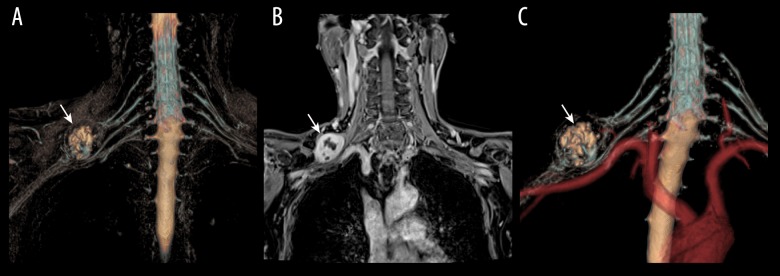
Figure 1.
Pathologically proven schwannoma of brachial plexus in a 40-year-old patient with right arm dysesthesia. (A) 3D T2-STIR-SPACE volume rending (VR) image of the brachial plexus revealed a round mass (arrow) in the upper trunk. (B) The tumors showed inhomogeneous contrast enhancement on the venous phase coronal VIBE image (arrows head). (C) 3D-synchro-view demonstrates the relationship of the mass to the surrounding vessels, and the subclavian artery was compressed and displaced (arrow).

