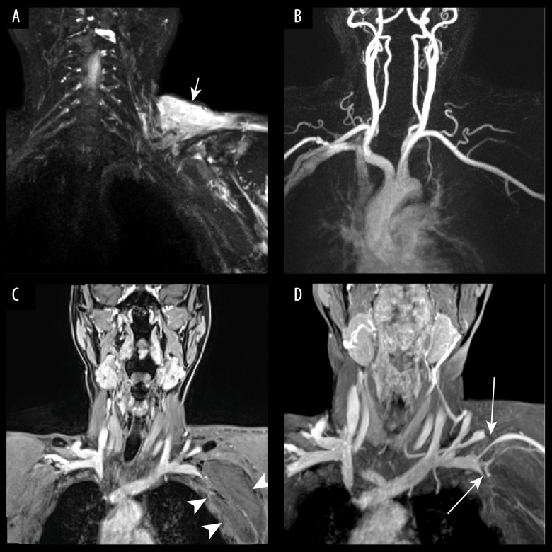
Figure 3.
A 72-year-old man with left arm swelling and finger numbness after shoulder trauma. (A) 3D-T2-STIR SPACE shows compression of left brachial plexus and wide-spread soft tissue edema of the left shoulder (arrow). (B) Contrast-enhanced MR angiography shows the left subclavian artery was normal. (C) Venous phase coronal VIBE image revealed a large axillary hematoma (arrows head). (D) Venous phase VIBE maximum-intensity-projection (MIP) image shows significant compression (arrows) of left subclavian veins (SCVs) and suprascapular vein.

