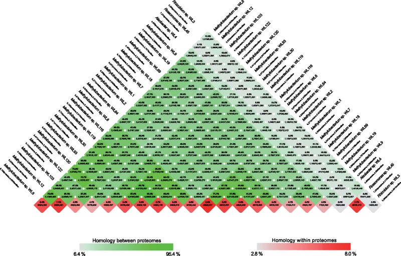
Fig. 3.
—Pairwise proteome comparisons (BLAST-matrix) of the 21 bacterial isolates of the analysis. The gray-to-green color gradient indicates low to high percentages of identity. The bottom boxes show the presence of homologs within the same proteome (gray-to-red color gradient shows low to high presence of homologs in the specific proteome).

