Figure 3.

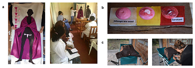
Depicting anatomy, pathophysiology, and the pelvic exam in a culturally sensitive manner. a. Anatomy was shown in situ superimposed on a human figure to help orient the women as we found that isolated pictures of organs in the human body which are “hidden” in the body did not register with the women. The uterus and cervix were discussed in terms of pregnancy and delivery as this is a familiar process to these women. b. The healthy cervix was shown alongside treatable pre-cancer and invasive cancer. The women held props made from doorknobs with painted clay “lesions,” which represented the progression from normal to invasive cervical cancer. c. A pelvic model made from felt and a cardboard box facilitated demonstration of the speculum exam. Women were also allowed to hold the speculum in order to demystify this tool.
