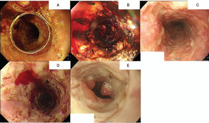
Figure 1.

Endoscopic features of a 55-years-old-male patient who was a heavy drinker. All endoscopic images were taken at the ER with a 3 to 6 months interval. At every visit, his chief complaint was hematemesis and black and white esophageal necrosis was discovered alternatively.
