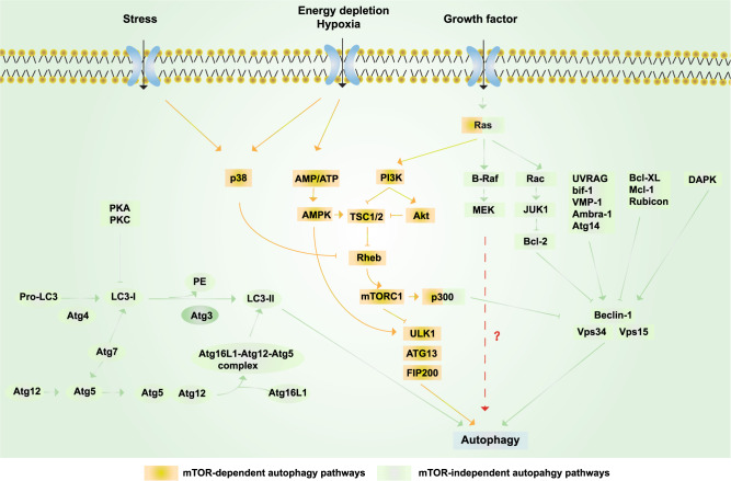
Fig. 4.
Schematic representation of autophagy. Yellow box: mTOR-dependent autophagy pathways. Growth factors can inhibit autophagy via activating the PI3K/Akt/mTORC1 pathway; under nutrient-rich conditions, mTORC1 is activated, whereas under starvation and oxidative stress, mTORC1 is inhibited. AMPK-dependent autophagy activation can be induced by starvation and hypoxia.449 Ras can also activate autophagy via activating PI3K,352 while p300 can inhibit autophagy.450 p38 promotes autophagy by phosphorylating and inactivating Rheb and then inhibiting mTOR under stress.451 Green boxes: mTOR-independent autophagy pathways. The PI3KCIII complex (also called the Beclin 1–Vps34–Vps15 complex) is essential for the induction of autophagy and is regulated by interacting proteins, such as the negative regulators Rubicon, Mcl-1, and Bcl-XL/Bcl-2, while proteins including UVRAG, Atg14, Bif-1, VMP-1, and Ambra-1 induce autophagy by binding Beclin 1 and Vps34 and promoting the activity of the PI3KCIII complex.357 In addition, various kinases also regulate autophagy. ERK and JNK-1 can phosphorylate Bcl-2, release its inhibition, and consequently induce autophagy; the phosphorylation of Beclin 1 by Akt inhibits autophagy, whereas the phosphorylation of Beclin 1 by DAPK promotes autophagy.452 Autophagy can be inhibited by the action of PKA and PKC on LC3. Finally, Atg4, Atg3, Atg7, and Atg10 are autophagy-related proteins that mediate the formation of the Atg12–Atg5–Atg16L1 complex and LC3-II.453 RAS and p300 can also regulate autophagy via the mTOR-independent pathway454

