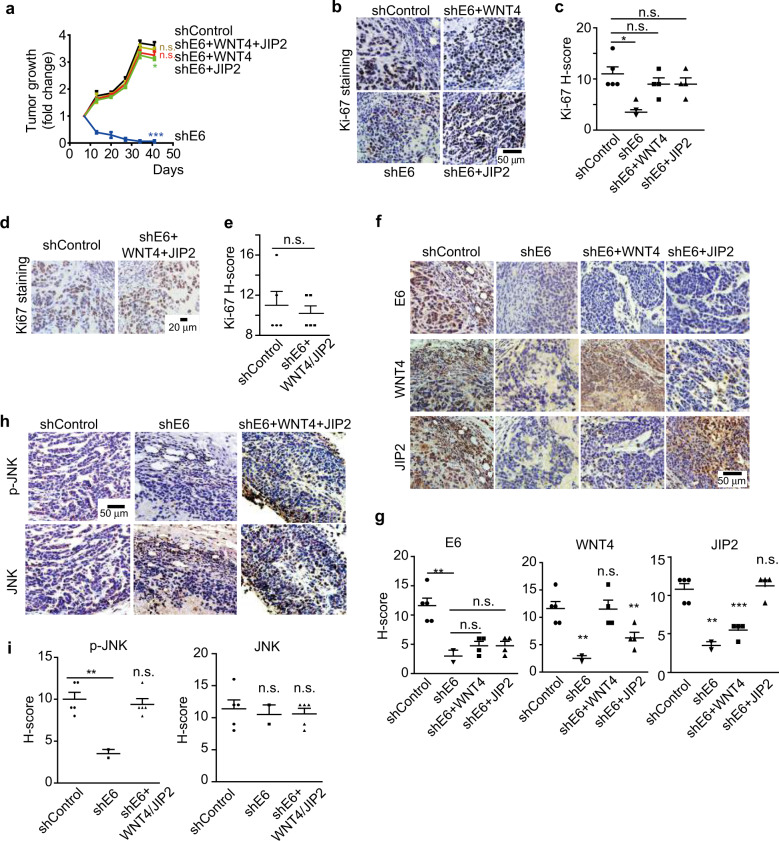
Fig. 4.
E6 promotes tumor growth through the activation of WNT/PCP/JNK pathway. a HeLa cells were stably transfected with the indicated shRNA or genes and injected subcutaneously into NOD/SCID mice. The growth of the tumor xenografts is plotted (n = 4–5). ‘n.s.’ denotes ‘not significant’, *p < 0.05. b–e Immunohistochemistry (IHC) staining for Ki-67 to show actively proliferating tumor cells in the xenografts. Representative images are shown in b and d. Quantitative results based on the H-score are plotted in c and e. ‘n.s.’ denotes ‘not significant’, *p < 0.05. f, g IHC staining of E6, WNT4, and JIP2 proteins in the indicated xenografts (f), and the protein levels were quantified based on the H-score (g). ‘n.s.’ denotes ‘not significant’, **p < 0.01, ***p < 0.001. h, i IHC staining for phosphorylated JNK (p-JNK) to show JNK activation in the indicated xenografts (h). The H-score-based quantification results are shown in i. ‘n.s.’ denotes ‘not significant’, **p < 0.01

