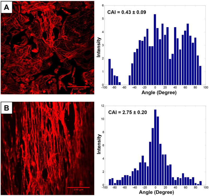
Figure 3.
Fluorescence imaging of hiPSC-CM labeled with phalloidin dye on a PLGA nanofiber scaffold. Cardiomyocytes seeded on the aligned nanofiber scaffold were aligned and elongated in the direction of the nanofiber, when compared to cells seeded on a flat cell culture plate. Alignment of cells on the scaffold was further confirmed by enhanced cell anisotropy index (CAI). Reprinted from Khan et al., PLOS One [121].

