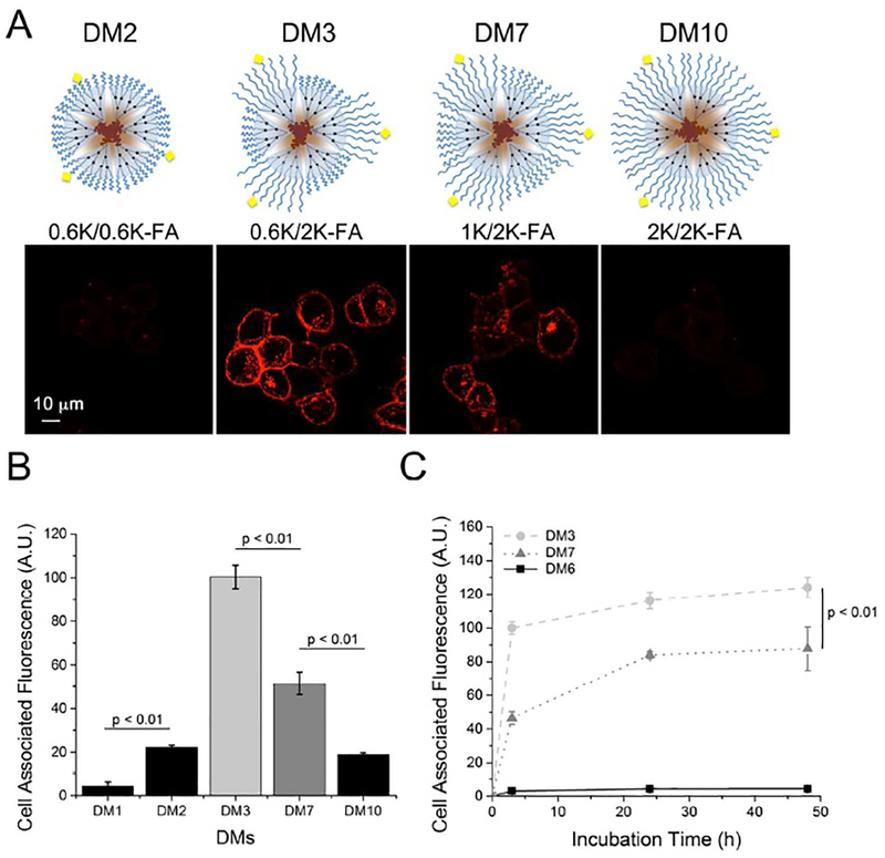
Figure 2.
Effect of PEG corona length on the FA-targeted cellular interactions and interaction kinetics of DMs. Each micelle contains 5 wt.% PDC2K-FA1. Confocal microscopy images of dendron micelles with varying PEG corona lengths (A) Quantification of DM cellular interactions by flow cytometry of various targeted DMs (B). Time-dependent FA-targeted cellular interactions of DMs, measured using flow cytometry (C).

