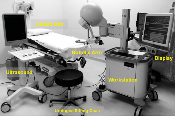
Figure 1.

PET-ultrasound fusion targeted biopsy system includes robotic arm, clinical ultrasound scanner and computer work station. Ultrasound probe was attached to robotic arm for 3-D ultrasound image acquisition. Prostate boundaries on 2-D ultrasound were segmented and used to generate 3-D prostate model. Computer work station was used to register PET-CT with 3-D ultrasound data. Lesion target seen on PET was mapped to 3-D model. PET-ultrasound fusion images were used to guide targeted biopsy in patients.
