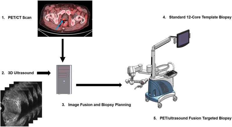
Figure 2.

Work flow and comparison of standard 12-core template biopsy with PET-ultrasound fusion targeted biopsy. 1, PET-CT images with fluciclovine acquired from patient show focal lesion in prostate (arrow). 2, prebiopsy 2-D ultrasound image was acquired in same patient using mechanically assisted navigation device and reconstructed into 3-D image. 3, 3-D ultrasound image was registered with PET-CT data to plan targeted biopsy. 4, at biopsy same mechanical device was used to acquire 2-D ultrasound images of patient. Computer generated 12-core template biopsy was performed under TRUS image guidance. 5, fused PET-ultrasound images were used to guide targeted biopsy while patient was on same platform.
