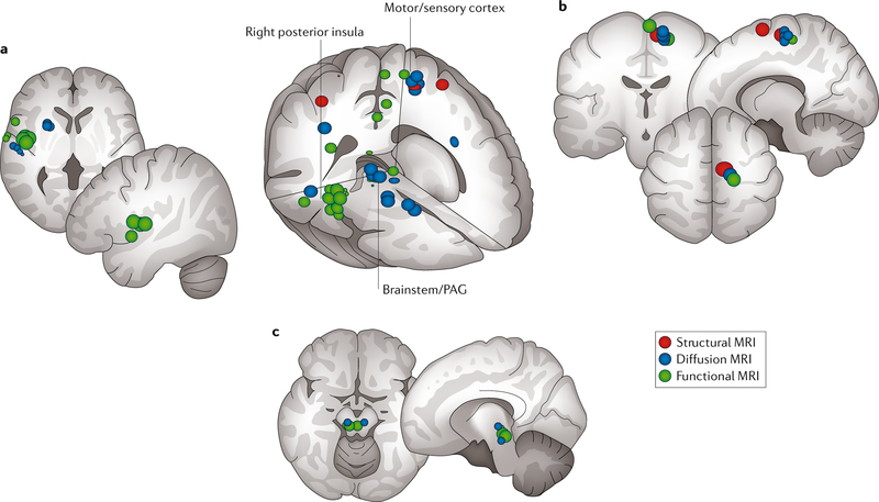
Fig. 3 |. MAPP I convergent neuroimaging findings.
Locations in the brain of altered function (green; assessed using resting-state functional MRI), grey matter structure (red; assessed using T1-weighted MRI) and white matter structure (blue; assessed using diffusion tensor MRI) in participants with urologic chronic pelvic pain syndrome (UCPPS) compared with matched healthy control individuals are shown51,52. We note particular overlap in the right posterior insula (part a), the medial sensorimotor areas (part b) and the brainstem and periaqueductal grey (PAG) (part c). MAPP, Multidisciplinary Approach to the Study of Chronic Pelvic Pain.

