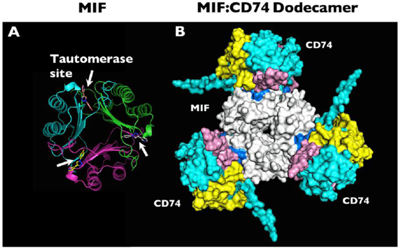
Figure 1.

(a) Molecular structure of MIF based on x-ray crystallography, with white arrows indicating the locations of the tautomerase sites between adjacent monomers. The tautomerase sites are shown occupied by the small molecule MIF20. (b) Computational model representation of the MIF trimer (white, center) engaging with CD74 trimers (blue, yellow, and pink, outer). Many small molecule MIF inhibitors can occupy the MIF tautomerase sites that appear in close apposition to the CD74 receptor. Reprinted by permission from Springer: Metabolic brain disease. Predicted structure of MIF/CD74 and RTL1000/CD74 complexes, Meza Romero R., et al, COPYRIGHT 2016.
