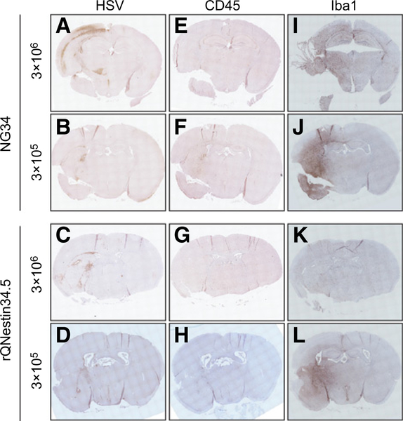
Figure 6.
IHC of the brains of athymic mice after the HSV-1 inoculation. Brains with 3 × 10⁶ pfu were obtained from the euthanized mice at a terminal point (day 3) during the toxicity study in Fig. 5C and D. Brains with the 3 × 10⁵ pfu were independently prepared for this study and obtained from the mice at day 5 after viral injection. The sections from the paraffin-embedded tissues were stained with anti-HSV1/2 (A–D), anti-CD45 (E–H), or anti-Iba1 (I–L) antibodies.

