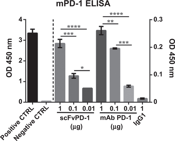
Figure 1.
In vitro evaluation of the ability of scFvPD-1 to bind PD-1. Specific interaction of the purified scFvPD-1 (scFv) and PD-1 mAbs (332.8H3) with murine PD-1 (mPD-1) was quantified by ELISA with a secondary antibody HRP-conjugated anti-myc or anti-mouse IgG1, respectively. Positive control (CTRL): coating with human c-myc peptide. Negative CTRL, coating with BSA. Absorbance (y-axis) represents optical density at 450 nm corrected by subtract reading at 570nm. Error bar shows the SEM. Group means were compared by one-way ANOVA (Tukey multiple comparisons test), where *, **, ***, and **** indicate P <0.05, 0.01, 0.001, and 0.0001, respectively (n = 3). The experiment shown is representative of experiments that were performed at least 3 times.

